CRANIAL CRUCIATE INJURIES IN DOGS ARE SOMEWHAT COMMON. WE PROVIDE THE SURGERY NECESSARY TO FIX THIS PAINFUL ISSUE.
What is the cranial cruciate ligament?
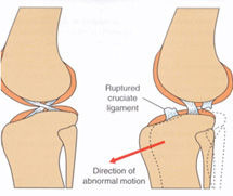
The cranial cruciate ligament (CCL) is a strong, rope-like structure inside the knee joint. It is the most important component of the knee as it constantly stabilizes the two bones involved in the joint. The CCL keeps the tibia (lower bone) from sliding forward and rotating internally while a dog is walking, running, or jumping. The femur (higher bone) also moves abnormally with each step when the ligament is injured, crushing the meniscus, a ligamentous “pad” that sits between the two bones. When the CCL is damaged and the knee is no longer stable, the abnormal movement of the bones is very painful. Over time, severe arthritis develops.
Cause
The cranial cruciate ligament in dogs is analogous to the anterior cruciate ligament (ACL) in humans. ACL tears are common in human athletes. In dogs, the ligament actually has to work harder due to the increased tibial angle relative to the ground. Newest research suggests cranial cruciate injuries in dogs occur gradually over a period of time even though these dogs exhibit pain acutely.
Diagnosis
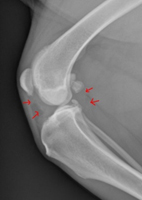
To diagnose a cranial cruciate injury, a few tests need to be done. The first is an orthopedic exam by the veterinarian. The veterinarian will try to pull the tibia forward. If they are able to do so, this is an indicator that the ligament may be ruptured. Radiographs (x-rays) of the knee are also important. The degree of disease within the joint can be evaluated as well as measurements taken of the slope of the tibia. Sometimes with partial tears of the ligament, we rely on the radiographs to validate our suspicion of cruciate disease. Sedation is also required to complete the orthopedic exam and procure adequate radiographs as the muscles of the leg need to be fully relaxed.
Non-Surgical Treatment
The primary treatment for a cruciate tear is surgical correction. Without surgery to stabilize the knee joint, the joint becomes arthritic. Scar tissue builds up around the knee, the body’s way of stabilizing the joint itself. Nonsurgical treatment involves strict rest and an anti-inflammatory medication for at least a few months. We will start the medication and recheck the patient in one month. The success rate of this conservative treatment is better in small dogs. At MLVH, we will recommend this treatment if surgical treatment is not financially feasible.
2012 © Synthes, Inc. or its affiliates. All rights reserved.
Surgical Treatment
With surgical correction and physical rehabilitation, the dog will feel much better and have improved use of the knee. Surgery lessens the arthritis development in the knee and helps relieve pain. This is especially important for younger dogs to give them higher quality of life.
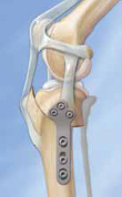
Tibial Plateau Leveling Osteotomy (TPLO) is one of the “bone-cutting” techniques and is designed to change the anatomy of the knee so that it no longer has abnormal movement of the bones. A semicircular cut is made at the top of the tibia with a curved saw so that the tibial joint surface is “leveled out”, to prevent forward slipping of the joint. A plate and screws are inserted to stabilize the cut bone during healing.
Lateral Suture Stabilization used to be the most common technique for surgical treatment of CCL disease. A suture (similar to fishing line) is placed around another ligament and into the tibia to provide stabilization of the joint during healing. The stabilization is temporary as the dog makes new scar tissue around the knee for long-term joint stability.
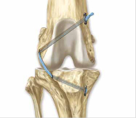
The TightRope procedure was developed in 2005 to provide a minimally invasive and improved method for stabilization of the joint. This technique does not require cutting of the bone like the TPLO. Instead, it uses two small, drilled tunnels in the femur and tibia to pass a synthetic ligament-like biomaterial. The biomaterial used for the TightRope is called FiberTape. It is a Kevlar-like material that is used extensively in human orthopedic surgery. The FiberTape acts as an artificial stabilizing ligament. As this material has very little stretch to it compared to the fishing line-type suture, the stabilization is more secure and lasts longer.
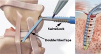
The SwiveLock Anchor procedure is very similar to the TightRope. FiberTape is used with this procedure as well. Instead of drilling two bone tunnels, only one is made. In replacement of the second bone tunnel, an orthopedic implant called a bone anchor is placed in the femur. This procedure is even less traumatic than the TightRope and a little faster to complete.
We offer both the SwiveLock Anchor and TightRope procedures at MLVH. The doctor will review which procedure is best for your pet.
Rehabilitation After Surgery
After surgical repair of the knee, a rehabilitation program is required to ensure faster recovery. We want your pet to be able to use their knee again! We have written out instructions for therapy at home, but professional rehabilitation is always encouraged. We can recommend a physical therapist in North Spokane (the closest to us). They will be able to use additional modalities such as an underwater treadmill, balance balls/boards, etc. Just let us know.
What's Next
Call us or schedule an appointment online.
Meet with a doctor for an initial exam.
Put a plan together for your pet.
